
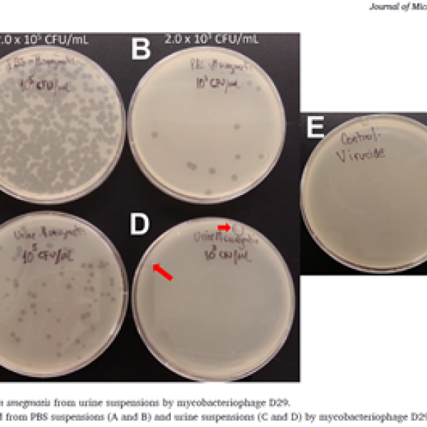
Figure. Detection of Mycobacterium smegmatis from urine suspensions using magnetic bead capture and mycobacteriophage D29 detection workflow. Reduced detectable signal at lower bacterial concentrations illustrates the operational sensitivity of specimen recovery workflows in urine matrices
Source: Adapted from Rojas-Ponce G et al. Journal of Microbiological Methods. 2022.
Operational Considerations in Urine-Based TB Diagnostics
Tuberculosis diagnostics continue evolving rapidly toward molecular testing, simplified workflows, and non-sputum-based approaches. Swab-based testing, oral sampling, urinary biomarkers, and molecular detection platforms are increasingly explored to improve access to diagnosis, particularly among children, people living with HIV, and patients unable to produce sputum.
This evolution raises an important question:
As new specimen types continue gaining attention, are we also paying enough attention to how these specimens are operationally processed before analysis?
Meanwhile, urine has been investigated for decades as a potential adjunct specimen for TB diagnosis. Historical reports describing urine as an adjunct specimen date back to the 1960s, and subsequent studies reported occasional recovery of Mycobacterium tuberculosis from urine specimens, including in some patients with pulmonary tuberculosis without overt genitourinary disease.
Despite this, urine has remained largely underused in routine TB laboratory workflows because of:
- low culture yield,
- inconsistent detection,
- contamination concerns,
- and methodological variability across studies.
As a result, most modern urine-based TB diagnostic development shifted toward:
- urinary LAM detection,
- transrenal DNA detection,
- and molecular assays.
However, an important operational question may still deserve attention.
Are current urine processing workflows fully optimized for recovery-sensitive specimens?
In many laboratories, urine continues to be processed using methodologies largely adapted from standard sputum decontamination and recovery workflows.
These typically include:
- centrifugation,
- pellet recovery,
- decontamination,
- buffer neutralization,
- re-centrifugation,
- resuspension,
- and inoculation into liquid or solid culture systems.
These workflows are standardized and operationally practical. However, important questions remain regarding their recovery efficiency when specimens may contain only very small numbers of bacilli.
For example:
- How efficient is centrifugation for recovering low numbers of viable bacilli from urine?
- How much variability exists between centrifuge performance, operators, and laboratories?
- Could pellet loss occur during decantation or resuspension?
- Could repeated centrifugation and decontamination steps contribute to cumulative bacillary loss?
- How much does residual alkalinity influence downstream recovery?
- And how much do processing variations influence molecular detection when only small aliquots are used?
These questions become particularly relevant when dealing with specimens that may contain very low concentrations of detectable material.
Biology versus recoverability
Current evidence strongly supports that:
- urinary LAM,
- transrenal DNA,
- and other mycobacterial components
may be detectable in urine under certain conditions.
At the same time, viable bacillary recovery remains inconsistent.
This raises an important distinction:
The biological presence of mycobacterial material may not necessarily correlate with efficient operational recoverability during laboratory processing.
In other words, low recovery may reflect:
- true biological scarcity,
- operational recovery limitations,
- or both.
Further methodological characterization may therefore be valuable before fully concluding the diagnostic limitations of urine as a specimen.
Experimental observations from urine recovery workflows
Previous experimental work exploring mycobacterial recovery from urine matrices highlights the potential operational sensitivity of low-burden specimen detection workflows.
Urine as a complementary specimen
This discussion is not intended to position urine as a replacement for sputum.
Rather, urine may continue to deserve attention as:
- a complementary specimen,
- a non-invasive specimen,
- and a potentially useful specimen in selected patient populations such as:
- people living with HIV,
- children,
- and patients with limited sputum production.
As diagnostic technologies continue evolving, optimizing specimen processing itself may become increasingly important.
Because diagnostic performance is not shaped only by technology.
It is also shaped by how specimens are operationally handled before analysis.
Moving forward
The objective of this discussion is not to replace sputum-based diagnosis, nor to oppose molecular innovation.
Rather, it is to encourage further reflection on whether specimen processing itself deserves greater scientific attention — particularly when dealing with specimens that may contain very small numbers of detectable organisms or targets.
As diagnostic technologies continue advancing, improving operational recovery and specimen handling may become just as important as improving analytical sensitivity itself.
In this context, urine may continue to deserve attention not only as a biomarker source, but also as a complementary specimen whose operational processing still requires further characterization.
Because sometimes, the challenge may not only be detecting the organism.
It may also be how we operationally recover it before analysis.
Selected References
- Colby FH. Essential urology. Baltimore: Williams and Wilkins; 1961. p. 552.
- Gopinath K, Singh S. Urine as an adjunct specimen for the diagnosis of active pulmonary tuberculosis. Int J Infect Dis. 2009;13(3):374-379.
- Peter J, Green C, Hoelscher M, et al. Urine for the diagnosis of tuberculosis: current approaches, clinical applicability, and new developments. Curr. Opin. Pulm. 2010; 16, 262–270.
- Chemeda A, Abebe T, Ameni G, et al. Utility of urine as a clinical specimen for the diagnosis of pulmonary tuberculosis in people living with HIV in Addis Ababa, Ethiopia. J Clin Tuberc Other Mycobact Dis.2019; 17:100125.
- Hikone M, Kondo Y, Takaizumi Y, et al. Urine-based diagnostic tests for tuberculosis: a scoping review highlighting unmet diagnostic needs. Front Microbiol. 2026. 17:1783312.
- Rojas-Ponce G, Sauvageau D, Zemp R, et al. Use of uncoated magnetic beads to capture Mycobacterium smegmatis and Mycobacterium avium paratuberculosis prior detection by mycobacteriophage D29 and real-time-PCR. J Microbiol Methods. 2022 Jun;197:106490.